#FITSurvivalGuide 1/10 Tweetorial on MR. From JASE April 2017. This is an extensive topic, and a tweetorial will not do justice, so highly recommend to read this article. @ASE360 @ACCCardioEd @newmexicoacc #echoboards 

2/10
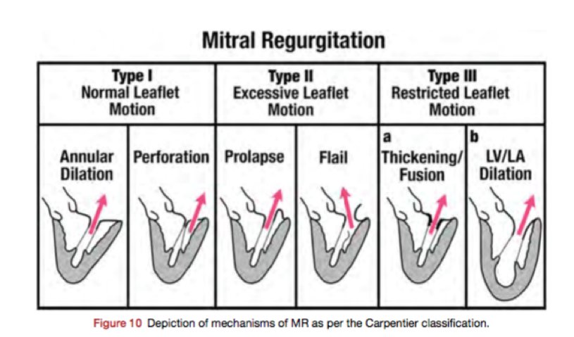
MR can be 1’ or 2’. Simplified version: 1’ MR d/t pathology of the valve itself. 2’ MR = MV apparatus is intact, w/ ventricular disease (LV dilatation) dilated MV annulus to MV leaflets malcoaptation MR (~central jet). Also useful to use Carpentier classification.
MR can be 1’ or 2’. Simplified version: 1’ MR d/t pathology of the valve itself. 2’ MR = MV apparatus is intact, w/ ventricular disease (LV dilatation) dilated MV annulus to MV leaflets malcoaptation MR (~central jet). Also useful to use Carpentier classification.

3/10
Determine the scallops. First heard from @MayoClinicCV: 2 steps
1.At 0 deg: Determine if ant or post leaflet abn
2.At 60 deg: Determine which scallop 1,2, or 3
Pic from J Am Soc Echocardiogr 2013;26:921-64
Determine the scallops. First heard from @MayoClinicCV: 2 steps
1.At 0 deg: Determine if ant or post leaflet abn
2.At 60 deg: Determine which scallop 1,2, or 3
Pic from J Am Soc Echocardiogr 2013;26:921-64

4/10
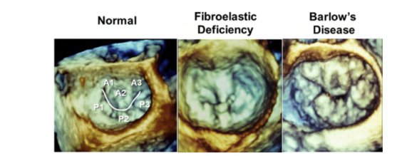
Can also use 3D, but be ware surgeon view vs TEE view (flipped). Here is the surgeon view, and others.
Can also use 3D, but be ware surgeon view vs TEE view (flipped). Here is the surgeon view, and others.

5/10
Severity of MR – extensive study. A few points here.
Vena Contracta (VC) – want to be perpendicular to flow when measuring. Should see flow converging to a narrow region (blue head and red head), before MR jet goes into the LA.
Severity of MR – extensive study. A few points here.
Vena Contracta (VC) – want to be perpendicular to flow when measuring. Should see flow converging to a narrow region (blue head and red head), before MR jet goes into the LA.

6/10
MR EROA measured by PISA radius. Blood approaches a narrow orifice, organizes into accelerating shells. Bring Nyquist baseline towards direction of jet (TTE Ap 4Ch view, move it up). Hemisphere surface area (SA)= 2∏r2. r = PISA radius. EROA = Alias Vel x SA / MR Peak Vel
MR EROA measured by PISA radius. Blood approaches a narrow orifice, organizes into accelerating shells. Bring Nyquist baseline towards direction of jet (TTE Ap 4Ch view, move it up). Hemisphere surface area (SA)= 2∏r2. r = PISA radius. EROA = Alias Vel x SA / MR Peak Vel

7/10
SV method can also be used to get RVol and RF. RVol = SV at Mitral Annulus – SV at LVOT. RF = RVol / SV at Mitral Annulus.
SV method can also be used to get RVol and RF. RVol = SV at Mitral Annulus – SV at LVOT. RF = RVol / SV at Mitral Annulus.

8/10
Important to keep in mind: if MR is holosystolic (HS) vs late systolic (LS) MVP. LS severe EROA ≥0.4cm2 MR may not be severe if the jet not going through it for the entire systole. In this case, base it off the RV. CW Doppler will show if HS vs LS.
Important to keep in mind: if MR is holosystolic (HS) vs late systolic (LS) MVP. LS severe EROA ≥0.4cm2 MR may not be severe if the jet not going through it for the entire systole. In this case, base it off the RV. CW Doppler will show if HS vs LS.

10/10
Again, not comprehensive. Hope you find this a good place to get started. Table for assessing severity attached here. Thanks to @dr_chirumamilla for the #FITSurvivalGuide - great idea! Glad to be part of it
Again, not comprehensive. Hope you find this a good place to get started. Table for assessing severity attached here. Thanks to @dr_chirumamilla for the #FITSurvivalGuide - great idea! Glad to be part of it

• • •
Missing some Tweet in this thread? You can try to
force a refresh


