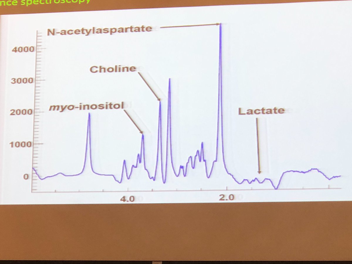
Jarred Younger up now. Neuroinflammation in #mecfs. We’ve all gotten inflammation diagnoses in the body (arthritis, e.g.) but never brain inflammation. Hard to diagnose! So how can we make that dx easier? (1/5) #pwme @MEActNet #MECFS18 

Hard to measure inflammation in a living person? Must be non-invasive, must be through and examine the whole brain. (2/5) #pwme @MEActNet #MECFS18
Microglia — bottom right is in an inflammatory state. Releases cytokines that cause ‘sickness behavior’. It makes you feel awful: that’s its job! Slow down to recover. (3/5) #pwme @MEActNet #MECFS18 

The issue is that microglia can get primed so that a short walk can trigger the immune response. “That’s what we think is happening.” (4/5) #pwme @MEActNet #MECFS18
SO GLAD he included the myo-inositol information. I researched this pathway at Stanford. Myo-inositol is also activated during encephalopathy. (5/5) #pwme @MEActNet #MECFS18 

Might be TOP RIGHT pls let's remember to check when livestream vid goes out!
• • •
Missing some Tweet in this thread? You can try to
force a refresh


