1/11
Today's #FiTSurvivalGuide for basic #EchoFirst views
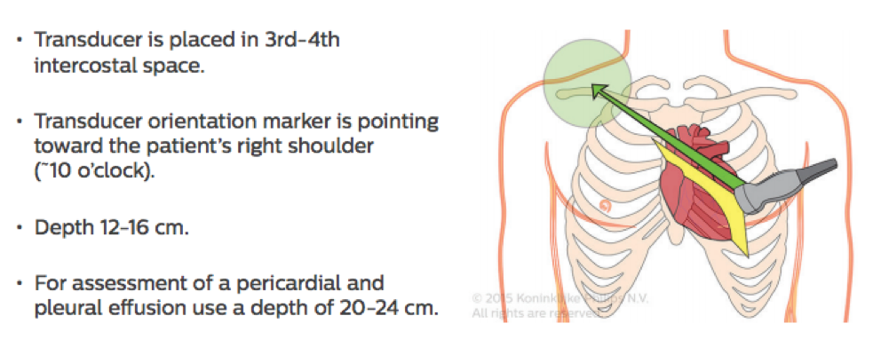
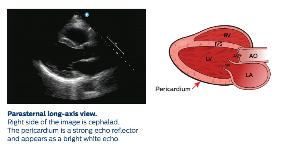
Parasternal Long Axis:
Left lateral decubitus
3rd L intercostal space. Move⬆️or⬇️ to find window
👀descending aorta, coronary sinus, pericardium, LV, both leaflets of MV, LA, aortic valve & root, RV

Today's #FiTSurvivalGuide for basic #EchoFirst views
Parasternal Long Axis:
Left lateral decubitus
3rd L intercostal space. Move⬆️or⬇️ to find window
👀descending aorta, coronary sinus, pericardium, LV, both leaflets of MV, LA, aortic valve & root, RV
2/11
RV Inflow View:
Medial angulation of scan plane
👀RA, Tricuspid valve, RV
Further angulate probe to remove portion of LV (seen in A, but not in B)
RV Inflow View:
Medial angulation of scan plane
👀RA, Tricuspid valve, RV
Further angulate probe to remove portion of LV (seen in A, but not in B)

3/11
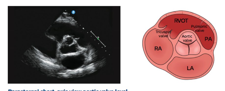
Parasternal short
👀annulus, 3 cusps of aortic valve (open in systole, close in diastole), coronary ostia (LM at 4 & RCA at 11), LA, IAS, RA, TV, RVOT, pulmonary valve, proximal pulmonary artery (slight superior angulation for R & L branches)
#FiTSurvivalGuide

Parasternal short
👀annulus, 3 cusps of aortic valve (open in systole, close in diastole), coronary ostia (LM at 4 & RCA at 11), LA, IAS, RA, TV, RVOT, pulmonary valve, proximal pulmonary artery (slight superior angulation for R & L branches)
#FiTSurvivalGuide
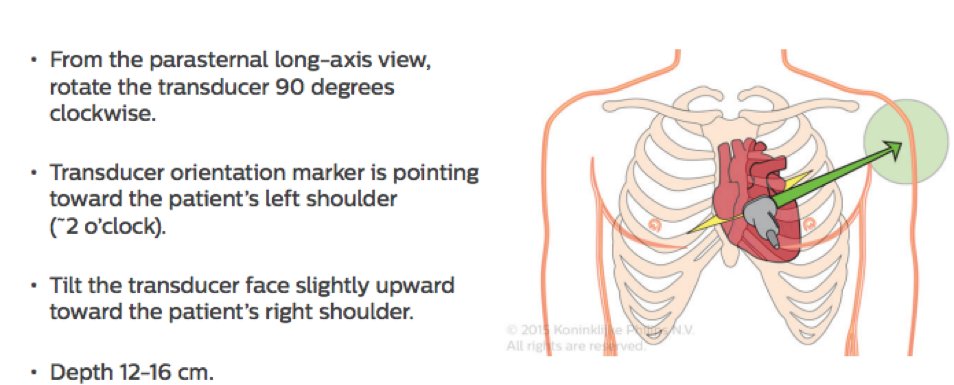
Rotate transducer slightly & adjust tilt ▶️ LV appears circular ▶️ both MV leaflets at maximal excursion
Visualize LV at: apex, papillary muscles, mitral valve
Visualize LV at: apex, papillary muscles, mitral valve

5/11
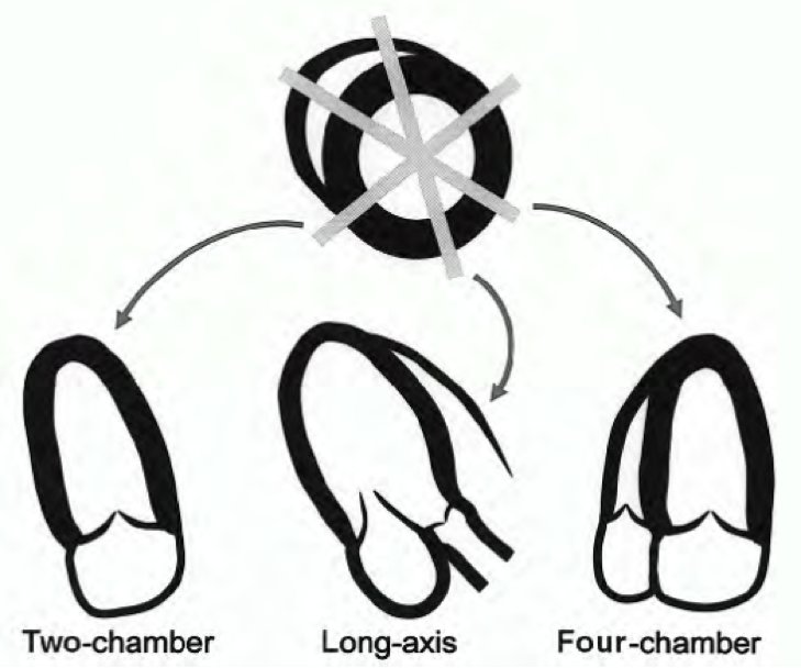
Apical views
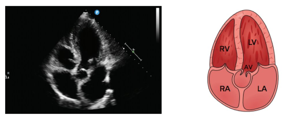
Apical 4 Chamber:
👀RA, TV, RV, LA, MV, LV & septa (IVS & IAS)
✔️TV septal leaflet insertion more apical than MV
✔️TV anterior leaflet is lateral
✔️Anterior MV is medial & Posterior leaflet is lateral

Apical views
Apical 4 Chamber:
👀RA, TV, RV, LA, MV, LV & septa (IVS & IAS)
✔️TV septal leaflet insertion more apical than MV
✔️TV anterior leaflet is lateral
✔️Anterior MV is medial & Posterior leaflet is lateral


6/11
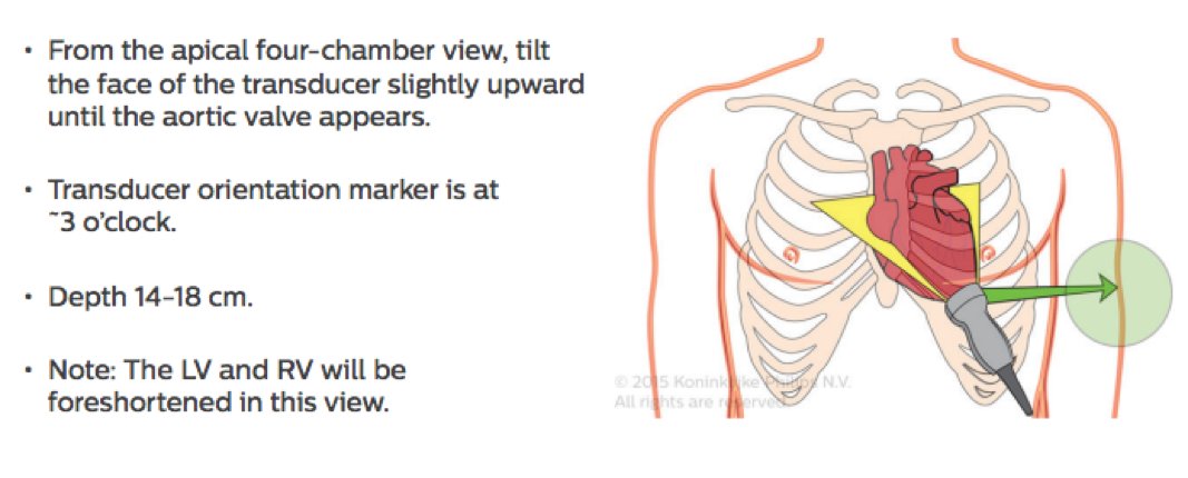
Obtain apical views via slight angulation and tilt of probe at same location.
Apical 5 Chamber:
5th "Chamber" is Aortic valve/LVOT
#FiTSurvivalGuide


Obtain apical views via slight angulation and tilt of probe at same location.
Apical 5 Chamber:
5th "Chamber" is Aortic valve/LVOT
#FiTSurvivalGuide
7/11
Apical 2 Chamber or Right Anterior Oblique equivalent:
Rotate counterclockwise 60 degrees
Exclude RA & RV; only visualize 👀 LV, LA, MV
✔️Sometimes can visualize left atrial appendage
Apical 2 Chamber or Right Anterior Oblique equivalent:
Rotate counterclockwise 60 degrees
Exclude RA & RV; only visualize 👀 LV, LA, MV
✔️Sometimes can visualize left atrial appendage

8/11
A3C or Apical Long Axis:
From A4C, rotate clockwise 60 degrees.
👀 MV and AV in same plane.
✔️Utility in detecting AV and subvalvular obstruction; HCM.
✔️May cause more endocardial dropout and poorer wall motion visualization.
#FiTSurvivalGuide
A3C or Apical Long Axis:
From A4C, rotate clockwise 60 degrees.
👀 MV and AV in same plane.
✔️Utility in detecting AV and subvalvular obstruction; HCM.
✔️May cause more endocardial dropout and poorer wall motion visualization.
#FiTSurvivalGuide

9/11
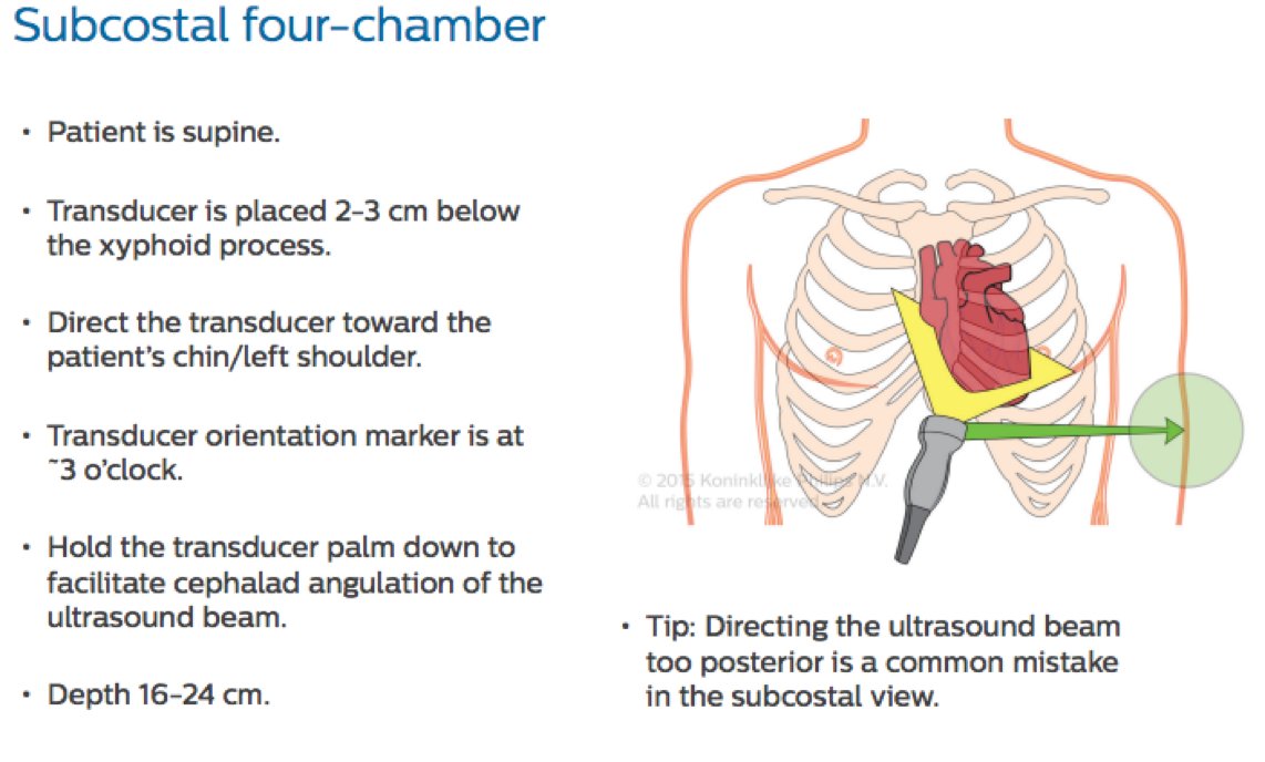
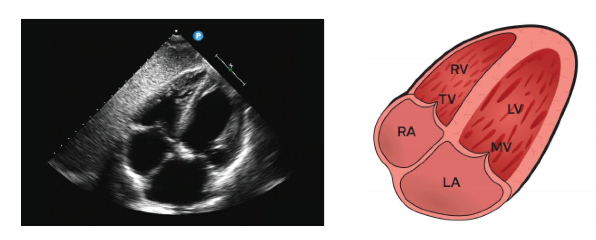
Subcostal 4 chamber; similar to apical view.
✔️Better endocardial definition
✔️Orientation of IAS and IVC ▶️ 👀septal defects; sinus venosus
✔️Assess RV free wall thickness & motion; pericardial tamponade ▶️ abnormal wall motion
✔️May foreshorten LV apex

Subcostal 4 chamber; similar to apical view.
✔️Better endocardial definition
✔️Orientation of IAS and IVC ▶️ 👀septal defects; sinus venosus
✔️Assess RV free wall thickness & motion; pericardial tamponade ▶️ abnormal wall motion
✔️May foreshorten LV apex
10/11
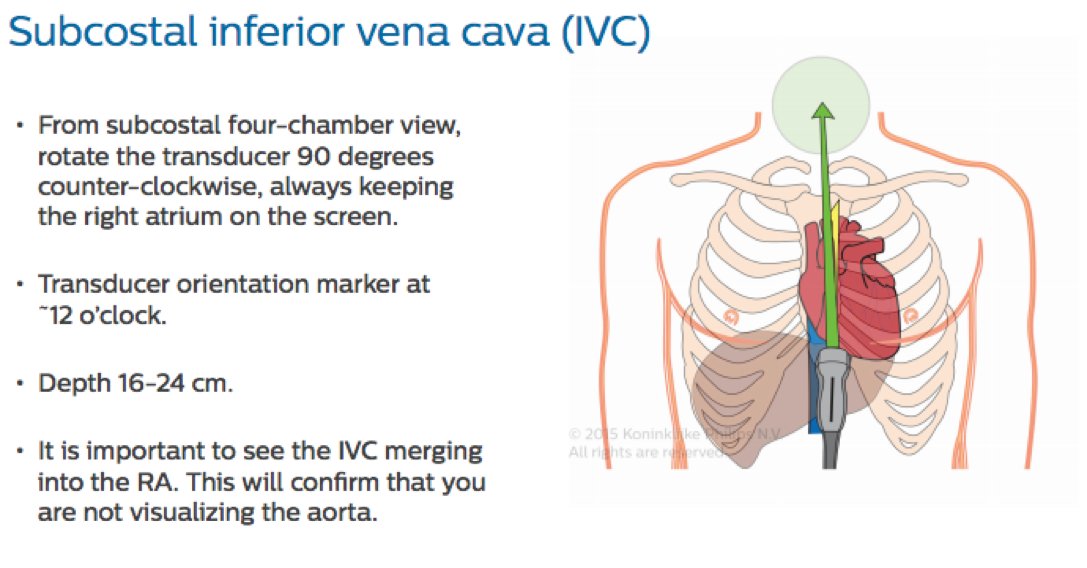
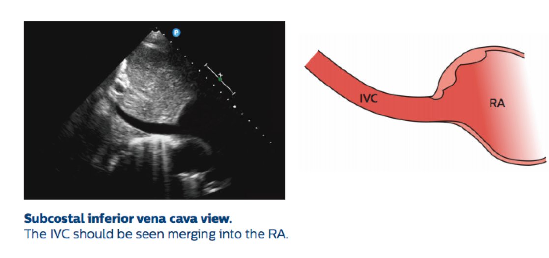
Subcostal IVC; via modification of short-axis plane
👀 IVC dimensions; sniff
✔️Hepatic vein flow; pulsed Doppler, especially with respiratory variation.
#FiTSurvivalGuide

Subcostal IVC; via modification of short-axis plane
👀 IVC dimensions; sniff
✔️Hepatic vein flow; pulsed Doppler, especially with respiratory variation.
#FiTSurvivalGuide
11/11
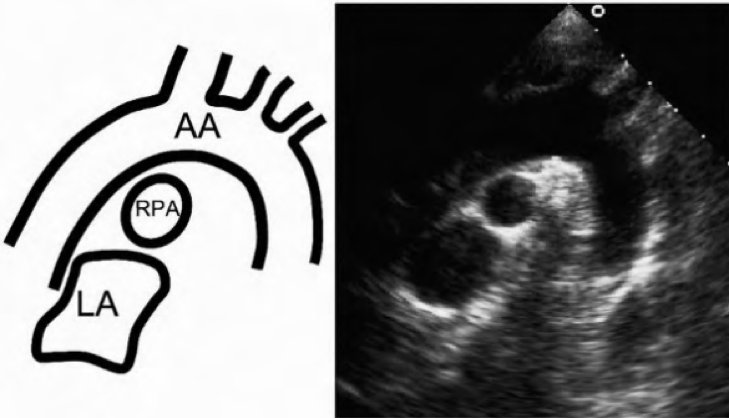
Suprasternal views: great vessels
Extend & rotate pt head
✔️Plane parallel to aortic arch; 👀ascending & descending aorta, origins of innominate, L Carotid, L Subclavian, R pulmonary artery
✔️90 degrees, perpendicular plane; 👀arch in short axis, L & R pulmonary artery

Suprasternal views: great vessels
Extend & rotate pt head
✔️Plane parallel to aortic arch; 👀ascending & descending aorta, origins of innominate, L Carotid, L Subclavian, R pulmonary artery
✔️90 degrees, perpendicular plane; 👀arch in short axis, L & R pulmonary artery

Here is the thread unrolled!
threadreaderapp.com/thread/1015216…
There is more #FITSurvivalGuide this month, especially with more #EchoFirst
threadreaderapp.com/thread/1015216…
There is more #FITSurvivalGuide this month, especially with more #EchoFirst
https://twitter.com/dr_chirumamilla/status/1015220084871135232?s=19
• • •
Missing some Tweet in this thread? You can try to
force a refresh


